Arteries Diagram : Arterial Supply Anatomy Overview Gross Anatomy Microscopic Anatomy - This is known as the main pulmonary artery or pulmonary trunk.
byAdmin-
0
Arteries Diagram : Arterial Supply Anatomy Overview Gross Anatomy Microscopic Anatomy - This is known as the main pulmonary artery or pulmonary trunk.. After receiving blood directly from the left ventricle of the heart, the. Inner body parts with their names. Check out our heart diagrams, quizzes and worksheets. The neck is supplied by arteries other than the carotids. The arteries' smaller branches are called arterioles and capillaries.
Check out our heart diagrams, quizzes and worksheets. The defect is associated with narrowing of the trachea (windpipe) and bronchi (airways). Is associated with venipuncture, it is done mainly by phlebotomists, nurses, emts and doctors. Some are more conceptual, others focus on branching, while still others attempt to preserve a spatial representation. An artery (plural arteries) (from greek ἀρτηρία (artēríā) 'windpipe, artery') is a blood vessel that takes blood away from the heart to one or more parts of the body (tissues, lungs, brain etc.).
Seer Training Classification Structure Of Blood Vessels from training.seer.cancer.gov To understand the system, the students need to create their diagrams. The aorta branches into a network of smaller arteries that extend throughout the body. An artery is an elastic blood vessel that transports blood away from the heart. Diagram showing the effects of atherosclerosis on an artery. Arteries and veins of the human body. The most common arteries diagram material is paper. The abdominal aorta bifurcates at the level of the fourth lumbar vertebra to form the two common iliac arteries, each of which further branches into the external and the internal iliac artery. Check out our heart diagrams, quizzes and worksheets.
Next, we have the blood vessel responsible for carrying deoxygenated blood from the right side of the heart (right ventricle) to the lungs.
Over the years, cholesterol plaques can narrow the arteries supplying blood to the heart. An artery (plural arteries) (from greek ἀρτηρία (artēríā) 'windpipe, artery') is a blood vessel that takes blood away from the heart to one or more parts of the body (tissues, lungs, brain etc.). Is associated with venipuncture, it is done mainly by phlebotomists, nurses, emts and doctors. Anatomy and function of the coronary arteries. The aorta branches into a network of smaller arteries that extend throughout the body. More artery diagrams are posted in the following 101 diagramss below. Arteries carry blood away from the heart in two distinct pathways: Arteries and veins of the human body. This is the opposite function of veins, which transport blood to the heart. Coronary arteries supply blood to the heart muscle. Inner body parts with their names. Some are more conceptual, others focus on branching, while still others attempt to preserve a spatial representation. Ascending aorta, aortic arch, thoracic aorta, and abdominal aorta.
Learn vocabulary, terms, and more with flashcards, games, and other study tools. The aorta branches into a network of smaller arteries that extend throughout the body. Learn the differences between an artery and a vein. The tunica medica, which is the very muscular middle layer in arteries, is thinner and less muscular in veins. The heart receives its own supply of blood from the coronary arteries.
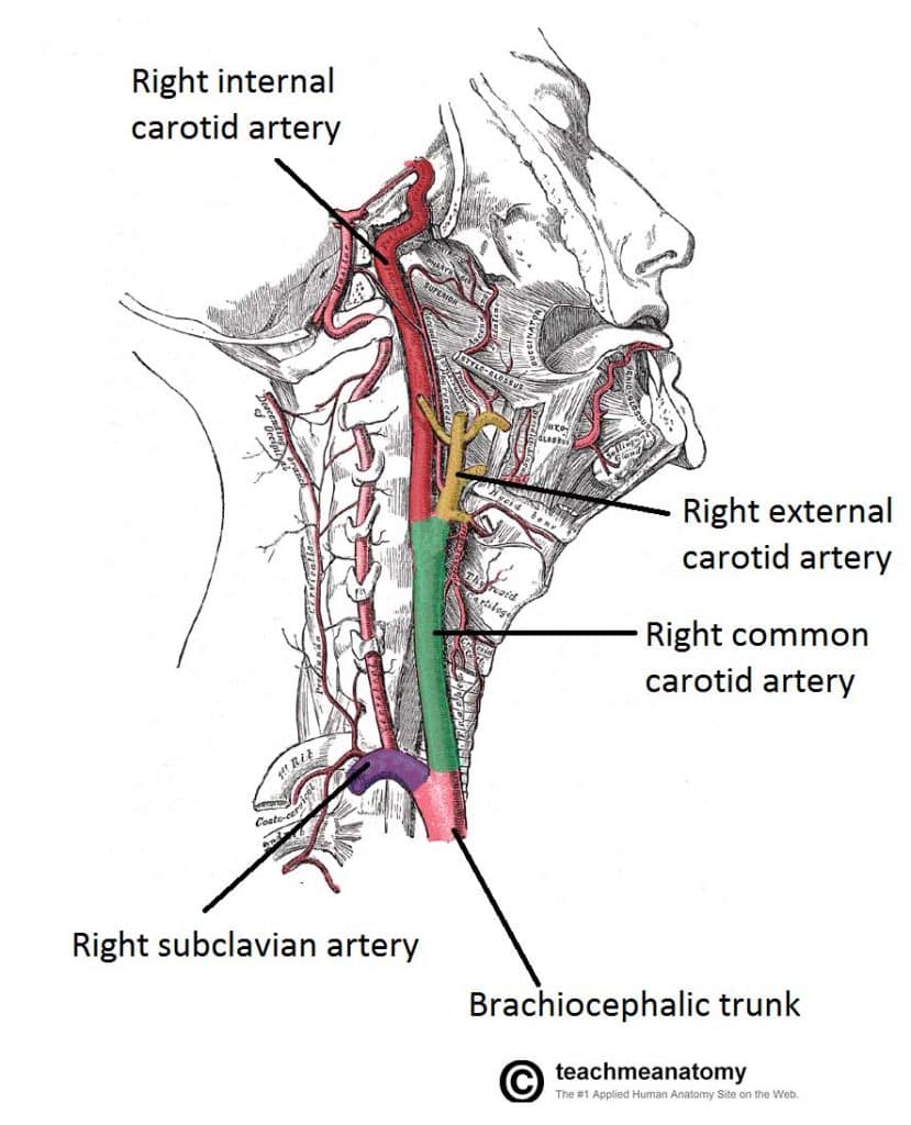
Spinal Arterial Anatomy Neuroangio Org from www.neuroangio.org Major arteries by definition, an artery is a vessel that conducts blood from the heart to the periphery. Check out our heart diagrams, quizzes and worksheets. Arteries and veins of the human body. Over the years, cholesterol plaques can narrow the arteries supplying blood to the heart. The most common arteries diagram material is paper. Arteries of the head and neck diagram art print vintage anatomy art print on tea stained paper dog art dog s wfh office art. In this image, you will find external carotid artery, internal carotid artery, vertebral artery, aorta and arch, pulmonary artery, cardiac artery, thoracic aorta, celiac trunk, superior mesenteric artery, renal artery, gonadal artery, inferior mesenteric artery, common iliac artery, external iliac artery. The cardiovascular system consists of the heart, blood vessels, and the approximately 5 liters of blood that the blood vessels transport.
These arteries and their branches supply all parts of the heart muscle with blood.
The main pulmonary artery splits into the right and left pulmonary arteries (better seen in the diagram at the end of this post). Blood is transported in arteries, veins and capillaries. 5 out of 5 stars. Labeled heart diagram showing the heart from anterior. The neck is supplied by arteries other than the carotids. The coronary arteries wrap around the outside of the heart. John bavosi/science photo library/getty images. Most arteries carry oxygenated blood; The narrowed arteries are at higher risk for complete blockage from a sudden. Create healthcare diagrams like this example called arteries and veins of the arm in minutes with smartdraw. It is returned to the heart in the veins. Blood is pumped from the heart in the arteries. Coronaryarteriescomplete from faculty.etsu.edu (taken from johnson, weipz and savage lab book).
Like maps, the various diagrams emphasize different aspects. Blood is pumped from the heart in the arteries. These vessels are channels that distribute blood to the body. Some are more conceptual, others focus on branching, while still others attempt to preserve a spatial representation. The veins also lack the elastic internal lamina that lies.
Major Arteries Of The Head And Neck Carotid Teachmeanatomy from teachmeanatomy.info Bodytomy provides a labeled iliac artery diagram to help you understand the anatomy and function of the common iliac. Next, we have the blood vessel responsible for carrying deoxygenated blood from the right side of the heart (right ventricle) to the lungs. Check out our heart diagrams, quizzes and worksheets. The neck is supplied by arteries other than the carotids. Blood is transported in arteries, veins and capillaries. Learn the differences between an artery and a vein. Two major coronary arteries branch off from the aorta near the point where the aorta and the left ventricle meet. Need a refresher on the basic anatomy of the heart?
The two exceptions are the pulmonary and the umbilical arteries, which carry deoxygenated blood to the organs that oxygenate it (lungs and placenta.
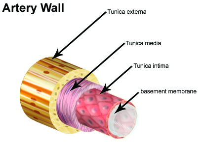
Each artery is a muscular tube lined by smooth tissue and has three layers: Two major coronary arteries branch off from the aorta near the point where the aorta and the left ventricle meet. It can also help them in getting an overview of artery vs. Human body artery diagram in detail. Like maps, the various diagrams emphasize different aspects. The right and left subclavian arteries give rise to the thyrocervical trunk. The first branch of the thyrocervical trunk is the inferior thyroid artery. Bodytomy provides a labeled iliac artery diagram to help you understand the anatomy and function of the common iliac. The heart receives its own supply of blood from the coronary arteries. Ascending aorta, aortic arch, thoracic aorta, and abdominal aorta. Learn vocabulary, terms, and more with flashcards, games, and other study tools. This is known as the main pulmonary artery or pulmonary trunk. 5 out of 5 stars.